This case is written by Dr. Donika Orlich. She is a staff physician practising in the Greater Toronto Area. She completed both her Emergency Medicine training and Clinician Educator Diploma at McMaster University.
Why it Matters
Many simulation cases that deal with pulmonary embolism seem to focus on the decision to administer thrombolytics (usually upon a patient’s arrest). This case is different. While the team must administer thrombolytics to a patient with known pulmonary embolism, the catch is that they must then also recognize shock as a result of intra-abdominal bleeding. As a result, the case highlights the following:
- The dose of thrombolytics to be used in the context of cardiac arrest
- The importance of an approach to undifferentiated shock after ROSC. (It’s not all cardiogenic!)
- That bleeding is a complication of thrombolysis. This is drilled into our brains as the major complication, but somehow it is diagnostically challenging to recognize.
Clinical Vignette
You are called urgently to the bedside of a patient who is in the Emergency Department awaiting medicine consultation. Your colleague saw her earlier. She is 63 years old and has a CT-confirmed pulmonary embolism. She had presented with shortness of breath on exertion in the context of a recent hysterectomy 4 weeks ago. She has been stable in the ED until she got up to go to the bathroom and suddenly developed severe shortness of breath.
Case Summary
A 63-year-old female is in the Emergency Department awaiting internal medicine consultation for a diagnosed pulmonary embolism. She suddenly becomes very short of breath while walking to the bathroom and the team is called to assess. The patent will then arrest, necessitating thrombolysis. After ROSC, she will stabilize briefly but then develop increasing vasopressor requirements. The team will need to work through the shock differential diagnosis and recognize free fluid in the abdomen as a complication of thrombolysis requiring surgical consultation and transfusion.
Download the case here: PE with Bleeding
ECG for the case found here:
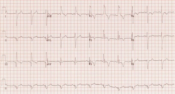
(ECG source: https://lifeinthefastlane.com/ecg-library/pulmonary-embolism/)
Initial CXR for the case found here:
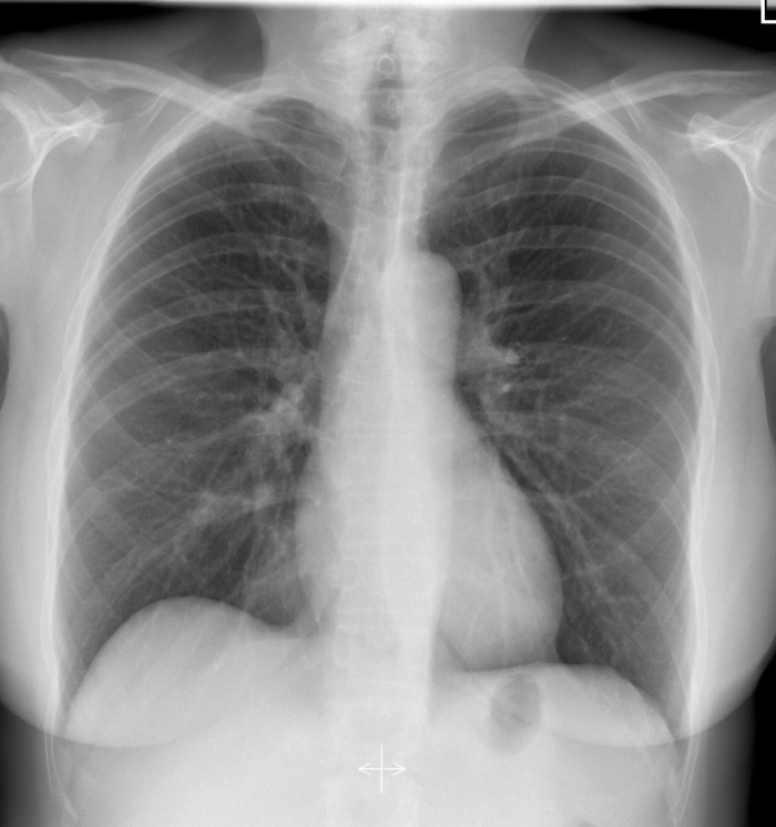
(CXR source: https://radiopaedia.org/cases/normal-chest-radiograph-female-1)
Post-intubation CXR for the case found here:
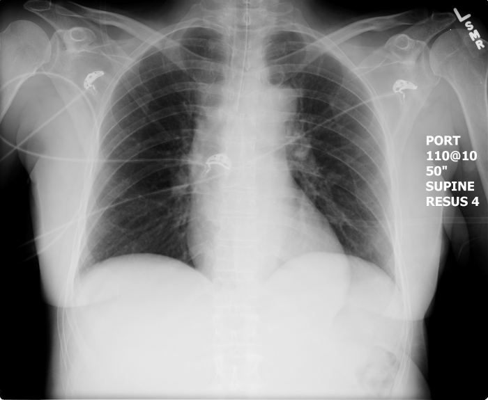
(CXR source: https://emcow.files.wordpress.com/2012/11/normal-intubation2.jpg)
Pericardial ultrasound for the case found here:
Normal lung ultrasound for the case found here:
Abdominal free fluid ultrasound for the case found here:

(All ultrasound images are courtesy of McMaster PoCUS Subspecialty Training Program.)
