This case is written by Dr. Donika Orlich. She is a PGY5 Emergency Medicine resident at McMaster University who also completed a fellowship in Simulation and Medical Education last year.
Why it Matters
While Emergency physicians certainly see their fair share of trauma, managing a patient with hemophilia is quite infrequent. This case highlights some key management points, including:
- The importance of administering early Factor VIII replacement
- The need to monitor for delayed intra-cranial hemorrhage
- The importance of determining capacity when a head-injured patient becomes agitated
Clinical Vignette
You are working in a level three trauma centre and are told that EMS just arrived from an MVC involving a 16-year-old female passenger who has known hemophilia. Vitals are stable. She has a laceration to her arm, and a bruise on her head, but has GCS 15 and only complains of arm pain.
Case Summary
A 16-year-old female presents following an MVC. Past medical history is significant for hemophilia A. She has a laceration on her arm and a bruise on her forehead, but denies HA/N/V. The learner should recognize high potential for bleeding, and implement immediate treatment with rVIII replacement, along with pan-CT imaging. The CT head will show a small ICH. The patient wants to leave AMA following normal CT results, and the learner must preform a capacity assessment and outline a plan of action for the incompetent patient. The patient should be sedated and/or intubated anticipating decline using neuroprotective measures. Consults should be made to the ICU and hematology.
Download the case here: Hemophilia Case
CXR for the case found here:
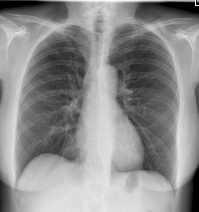
(CXR source: https://radiopaedia.org/cases/normal-chest-radiograph-female-1)
PXR for the case found here:
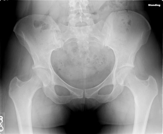
(PXR source: http://radiopaedia.org/articles/pelvis-1)
Forearm x-ray for the case found here:

(X-ray source: http://www.auntminnie.com/index.aspx?sec=ser&sub=def&pag=dis&ItemID=56736)
ECG for the case found here:

(ECG source: https://lifeinthefastlane.com/ecg-library/sinus-tachycardia/)
FAST image for the case found here:
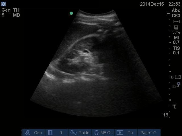
Cardiac U/S showing no pericardial effusion found here:
(U/S images courtesy of the McMaster PoCUS Subspecialty Training Program)
