This case is written by Dr. Kyla Caners. She is an emergency physician in Hamilton, Ontario and the Simulation Director of McMaster University’s FRCP-EM program. She is also one of the Editors-in-Chief here at EmSimCases.
Why it Matters
Pancreatitis is a common diagnosis made in the ED. However, severe pancreatitis with shock is relatively rare. As such, this case highlights several important points about the management of a hypotensive patient with abdominal pain:
- The importance of maintaining a broad differential diagnosis and employing beside imaging in one’s assessment
- The need for aggressive fluid resuscitation in an acutely hypotensive patient
- The risk of ARDS with pancreatitis
- The importance of developing a safe approach to the intubation of a patient who is simultaneously hypoxic and hypotensive
Clinical Vignette
Patricia is a 50 year old female who presents with epigastric abdominal pain. It’s been persistent for the last 24 hours and radiates through to her back. She has been nauseous all day and has been vomiting so much she “can’t keep anything down.” She was “on a bender” this weekend drinking beer and whiskey.
Case Summary
A 50 year-old female who was “on a bender” over the weekend now presents with diffuse abdominal pain and persistent nausea and vomiting. She will have a diffusely tender abdomen, a BP of 80/40, and be tachycardic. The team will need to work through a broad differential diagnosis and should fluid resuscitate aggressively. Once the patient has received 6L of fluid, she will become tachypneic and hypoxic and require intubation. The team will be given a lipase result just prior.
Download the case here: Pancreatitis with ARDS
ECG for the case found here:

(ECG source: http://cdn.lifeinthefastlane.com/wp-content/uploads/2011/12/sinus-tachycardia.jpg)
Initial CXR for the case found here:
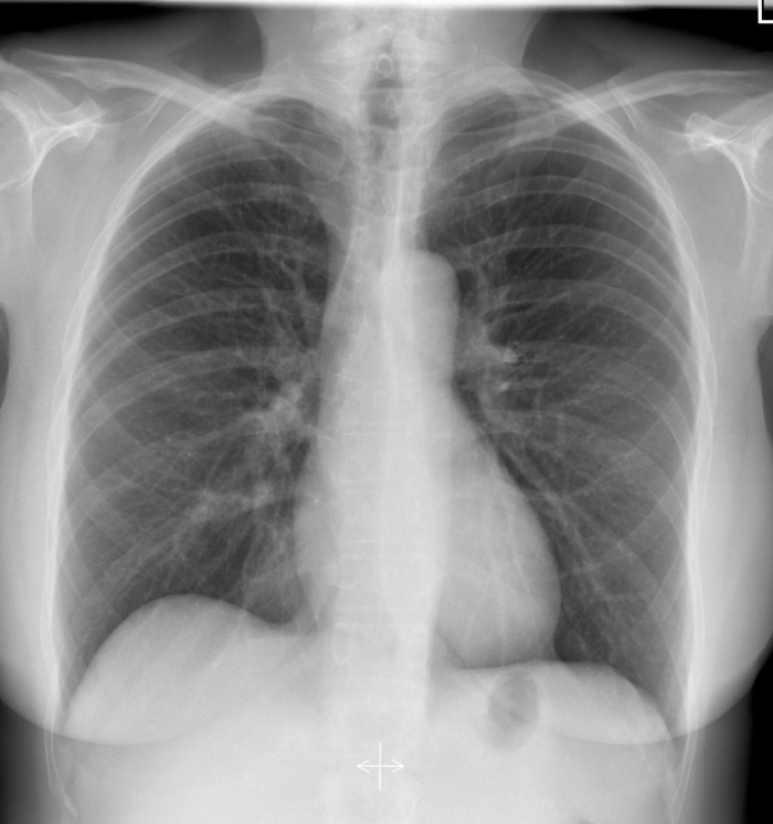
(CXR source: http://radiopaedia.org/articles/normal-position-of-diaphragms-on-chest-radiography)
ARDS CXR for when patient is hypoxic found here:
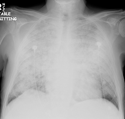
(CXR source: http://www.radiology.vcu.edu/programs/residents/quiz/pulm_ cotw/PulmonConf/09-03-04/68yM%2008-03-04%20CXR.jpg)
Post-intubation CXR for the case found here:
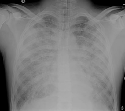
(CXR source: http://courses.washington.edu/med620/images/mv_c3fig1.jpg)
FAST showing no free fluid found here:
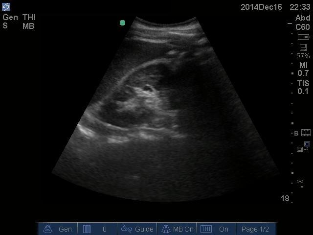
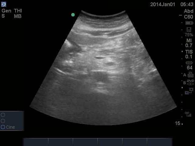
U/S aorta showing no AAA found here:
Pericardial U/S showing no effusion found here:
(All U/S images are courtesy of McMaster PoCUS Subspecialty Training Program)
