This case is written by Dr. Kyla Caners. She is a staff emergency physician in Hamilton, Ontario and the Simulation Director of McMaster University’s FRCP-EM program. She is also one of the Editors-in-Chief here at EmSimCases.
Why it Matters
Management of trauma patients with multiple intercurrent injuries can be challenging. This case provides an opportunity for junior learners to stretch themselves beyond their comfort zones. In particular, this case highlights the following:
- The need for a systematic approach to the initial assessment and ongoing re-assessment of any complex trauma patient
- The importance of prioritizing tasks and adjusting priorities as patient status changes
- The complexity of managing a hypotensive, head-injured patient
Clinical Vignette
A 32-year-old female presents as a trauma activation with EMS after being bucked off of her horse. Her mom witnessed the episode and called EMS because she seemed groggy. She has had a low BP with EMS on route. Her current BP is 80/40.
Case Summary
A 32-year-old female presents after being bucked off of her horse. She is brought in as a trauma team activation because of a low BP. Her primary survey will reveal a boggy hematoma over her right temporal area as well as an unstable pelvis. Her initial GCS will be 8. The team will proceed through airway management in a hypotensive, head-injured trauma patient while also binding her pelvis. The patient eventually shows signs of brain herniation, which the team will need to manage prior to consultant arrival.
Download the case here: Pelvic Fracture and SDH
ECG for the case found here:

(ECG source: https://i0.wp.com/lifeinthefastlane.com/wp-content/uploads/2011/12/sinus-tachycardia.jpg)
Pre-intubation CXR for the case found here:
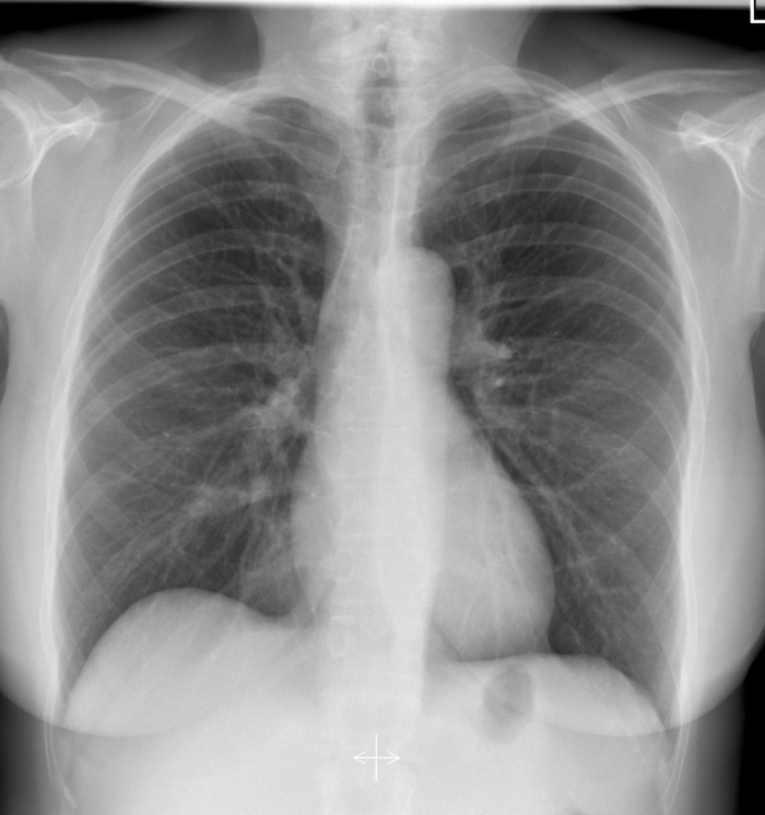
(CXR source: https://radiopaedia.org/cases/normal-chest-radiograph-female-1)
PXR for the case found here:

(PXR source: https://littlemedic.files.wordpress.com/2013/01/pelvis_0_1.jpg)
Post-intubation CXR for the case found here:
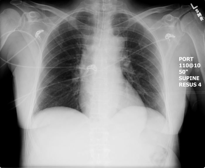
(CXR source: https://emcow.files.wordpress.com/2012/11/normal-intubation2.jpg)
Ultrasound showing free fluid in RUQ found here:

Ultrasound showing normal lung sliding found here:
Ultrasound showing no pericardial effusion found here:
(All U/S images are courtesy of McMaster PoCUS Subspecialty Training Program)
